The 2023 Submissions & Results

We received 28 fine photos for the image award this year.

The results from the judges:

FIRST PLACE: Khylan Stubblefield- monsterra plant
SECOND PLACE: Alex Thomas- iguana
THIRD PLACE: Ashley Wong ’24- polyp

Thank you to the judges and to all of the students who entered such lovely images!

1-Grant Nguyen:
King of the Savannah: The Alpha Lion of the Mabula Game Reserve. This photo was taken during the Men’s Soccer Team’s morning safari in Bela-Bela, South Africa. The team traveled to South Africa in August 2022, where they encountered the alpha lion and his pride of lionesses. Along with many other unique animals that also included elephants, rhinoceros, zebras, cheetahs, the Mabula Game Reserve is home to a plethora of wildlife, many of which are endangered, making these real-life sightings even more fleeting and their protection and preservation of the utmost importance. 
2-Winnie Lin:
This is an image of a polyp of hydractinia studied in my Stem Cell Biology course. The bright green dots (RFamide::GFP) show neuron activity!
3-Adrianna Belskis ’24:
Echinacea purpurea, the purple coneflower, photographed at Tyler Arboretum in March 2022. Observe a powerful pollinator, the eastern bumble bee (bombus impatiens), enjoying a peaceful afternoon amid the purple coneflower patch. 
4-Sharon Liu:
Anemone tentacle observed under a microscope for Bio 2 lab.  Fun fact: there are many dinoflagellates, a single-called organism, that lives within an anemone in a symbiosis relationship. The dinoflagellates photosynthesize and provide energy for its host anemone!
5-Alex Thomas: SECOND PLACE
Unimpressed by the tourists; Galapagos Yellow Land Iguana, Conolophus subcristatus, taking in the rays at sunset in the grass on Santiago Island.
6-Chenhao Xu:
I captured the moment of the cicada breaking out of its shell and gaining its wings after years of development under the ground.
7-Jasmine Bao ’24:
Aboral surface of a live Pacific purple sea urchin (Strongylocentrotus purpuratus), taken under a dissecting microscope in the Animal Biodiversity lab. Sex determination in sea urchins is usually achieved through identifying the type of gamete released from their aboral gonopores, with yellow being eggs and white being sperm, as sea urchins lack external sexual dimorphism. In addition to mineralized spines on the body surface, sea urchins also have thin, small tube feet (pictured) driven by a water-vascular system that aid in feeding, locomotion, and circulation.
8-Mina Mandic:
This is a microscope image of liquid crystal defects under a polarized optical microscope. Liquid crystals are a state of matter in between solid and liquid, so the molecules usually point in the same direction but they can also interact with each other and twist! A defect is a spot at which the two different twisting directions (clockwise and counter-clockwise) meet. Visualizing and understanding liquid crystal defects has many applications in biology, technology, and physics!
9-Evelyn Parts:
This photo, taken in my hometown of Towson, Maryland, captures a juvenile Cooper’s Hawk perched on a branch after a meal. His bright yellow eyes and lack of a dark gray cap are indicative of a juvenile male. To fight off the cold, his one foot is nestled in his belly feathers. As I watched him, he would switch which foot he held onto the branch with every so often. At the base of his beak, there is a little bit of a blood stain from his last meal. I often see him around my neighborhood and was really excited to get this shot.
10-Jesus Saucedo Bucio ’26:
This is a picture of a Drosophila willistoni fruit fly. The picture was taken through a microscope when looking for physical differences between diverged species of Drosophila in BIOL 002 Lab.
11-Ashley Wong ’24: THIRD PLACE
Sometimes it’s just not a match! Here is one twirling Hydractinia feeding polyp under transmitted light using fluorescence microscopy. After a one month recovery, the Hydractinia polyp rejected transplanting with an additional polyp.
12-Jonah Ring: HONORABLE MENTION
Two Great Tiger Moths (arcatia Caja) found near the peak of Mt. Ida in Northern Colorado hunker down against the harsh alpine winds. The vibrant coloration on these moths wards off potential predators; their body fluids contain neurotoxic choline esters.
13-Halle McLean:
Title: Dogwood at Dusk. The big “flowers” of the flowering dogwood, or Cornus florida, aren’t quite as they appear. What most interpret as large “petals” are actually bracts––or modified leaves; in most varieties these are white, but pink and red variations have also been found. The true flowers are the inconspicuous green beads at the center of these leaves, and when fertilized, they develop bright red fruits. As a bisexual flower, the dogwood is capable of self-fertilizing. Overall, dogwoods are important to wildlife and people alike: the plant is a food source for caterpillars and birds, but farmers also use the blooming of dogwoods to signify the true start of spring, waiting to plant their crops until they receive this signal.
14-Johanna Lee:
Crossvine (Bignonia capreolata) blooming along the exterior of Wharton Hall in late April. This vine can grow to be 50 ft. long and is frequented by hummingbirds. 
15-Nour Alajarma:
Swarthmore College in spring: tree in full bloom
16-Sander Simon: For a Biology project my group went out and explored the Swarthmore campus identifying plant species. We saw the beautiful lane of dawn redwoods by LANG and got this great photo of the afternoon light through the leaves (or leaflets!) of the trees!
17-Ana Carney ’24:
This photo was taken one morning during a semester abroad in a town called Maceda in Galicia, Spain. Maceda is a small town in Galicia with ~3,000 inhabitants and many natural landscapes. A small black horse can be seen grazing in a patch of grass in front of a distant mountain range.
18-Kennedy Hill: This membrane filter was part of a project that studied the removal of E. coli from Crum Creek water using Pearl Oyster and Turkey Tail Mushrooms. A sample of treated Crum water was filtered through this membrane. After incubating for 24 hours, E. coli colonies (yellow) were counted.
19-Ava Golde: Taken on a morning walk in Crum woods, this photo captures a series of fungi growing on a fallen log. Scattered across the tree, they give the impression of a series of shells found on a beach. Seen from far away, they don’t look like much, but up close showcase a beautiful gradient of neutral tones.
20-Aviva Weiser: (no caption)
21-Katelynn Swaim: This is a greater white-fronted goose nest with 9 eggs located on the coastal plains of the Yukon-Kuskokwim Delta in western Alaska. Each year these Arctic-nesting geese typically lay between 4 and 7 eggs in large insulated nests. This nest was observed as part of the 2022 Yukon-Kuskokwim Delta Nesting Study. 
22-Ainsley Jane Tambling: A tree peony (paeonia zeus) found outside the intercultural center at Swarthmore College. Their blooms can grow up to the size of a dinner plate and only last for around a week. They can be found throughout the eastern, midwestern and western United States. 
23-Ty Orsag: A Coral-Pink Merulius that I found growing on a fallen, rotting tree deep in the Crum Woods. This fungi, like most fungi, is a saprobe, breaking down dead organisms for nutrients.
24-Khylan Stubblefield: FIRST PLACE
Early one spring morning a glistening Monsterra plant is illuminated by the warm glow of the Swarthmore College greenhouse as the plants are misted.
25-Ruby Novogrodsky ’25: A clump of Dicranum moss hangs out on the bark of a tree at Olympic National Park. The tiny brown sporophytes, whose cells will divide via meiosis to produce spores, reach for the sun on green setas, or stalks. Soon, those spores will disperse to grow into new clumps of moss elsewhere.
26-Caleb Scott-Joseph: The Spotted Newt is a common sight in the Eastern United States with its very noticeable warning coloration. I found this one while on a walk in the woods of western Virginia near the Mountain Lake Biological Station. The orange Newts are actually the juvenile stage of this species. When it becomes an adult it will become brown in color and become exclusively aquatic.
27-Lane Barron: A flowering tree peony welcomes in a bee to engage in pollination, an act of mutualism.
28-Lina Verghese ’25-This is an image of a crayfish’s tail that was positioned under a brightfield microscope. In Bio 022, we dissected the tail to expose the dorsal part of the abdomen to measure neuronal activity in living tissue through mechanical stimulation of muscle stretching using an electrode.

The 2022 Submissions & Results

We received 22 fine photos for the image award this year.

The results from the judges:

FIRST PLACE: Ruby Novogrodsky ‘25- Star Wars magnolia flower
SECOND PLACE: Tristan Walker-Andrews- Caribou
THIRD PLACE: Alyssa Hayashi ’25- Anemone
HONORABLE MENTION: Jonah Ring ’23- Coral mushroom

Thank you to the judges and to all of the students who entered such lovely images!


1-Momi Jeschke: The beautiful early purple crocus (Crocus tommasinianus) gather to bloom together near Wharton dormitory in March, 2021, marking the true arrival of spring. 


2-Daniel Oakes ’24: One of the Scaphiopus couchii (Couch’s spadefoot toad) in the Alexander Baugh lab. This was when we were rehoming them into their new tanks and this female (affectionately named frogman) was posing up a storm. Spadefoot toads are so named because they have a little keratinized spade on their hind legs that they use to dig themselves into whatever substrate they live in.


3-TJorge Padilla ’24: Testing out my macro camera to capture very close up but high def. images, and I was very happy with how this photograph turned out. I find it cool how the fake background of the zoo enclosure looks real in this picture, and how the turtle looked into the camera as if knowing it was being photographed. I also feel like there’s a bit of a dramatic, evocative essence to the picture. The turtle’s glare almost looks as if it’s calling for humanity to heed the lectures of Mother Nature and treat our environment with more respect and care.

4-Fatima Jahra ’24:
“I’m just a sea-anemone posing for a photoshoot, while I’m under a compound microscope in the bio lab! The biologists here today are trying to figure out whether the symbionts in my body cause me to respond to light.”

THIRD PLACE WINNER: 5-Alyssa Hayashi ’25:
This is an Aiptasia pallida sea anemone with a close up of its mouth, oral disk, and tentacles. In my Bio 2 lab, we were identifying the role that the symbiont, Symbiodinium, had on the sea anemone’s behavior in response to a light source. 

6-Emily Kerimian:
This photo features a robin perched on a tree. Robins are common in the Crum, as well as across Swarthmore’s campus. Robins tend to be most active during the winter months. I took this picture near Wharton, where there are many pine trees that robins frequent. As a member of bird club and general bird enthusiast, I am always on the lookout for a subject, and this robin proved to be very photogenic.

7-Julia Stern:
This image shows an  American Eastern Yellow-Orange Fly Agaric Mushroom (Amanita muscaria var. guessowii) photographed on the coast of Eastham Pond in New Hampshire in August of 2021. I named her Maeve the Mushroom:).

HONORABLE MENTION- 8-Jonah Ring ’23:
This yellow-tipped coral mushroom (Ramaria Formosa) glows luminously in the afternoon sunlight of the Mount Zirkel Wilderness in Northwestern Colorado. R. Formosa has been shown to have mycorrhizal associations with conifers in the Western U.S.; the specimen shown here was found amidst a grove of pines and spruces.

9-Sophia Schlenz:
Gold Cobblestone Lichen (Pleopsidium flavum) and Elegant Sunburst Lichen (Xanthoria elegans) soaking in the sun on the high Sierra Nevada mountains in California. This photo was captured while I was exploring a dry alpine ecosystem during a visit home and features a variety of crustose lichens that thrive at an elevation of around 10,000 ft. Lichens are composite organisms that are the result of a unique symbiosis between fungi and either alga or cyanobacterium. Crustose lichen can live for thousands of years and is found nearly everywhere on our planet, from Antarctica to deserts, and alpine peaks like these. Map lichens are even considered Earth’s oldest organism, making lichens even cooler than they already were.

10-Olivia Fey ’23:
 A single, bisexual fern gametophyte viewed under a compound scope at 100 micrometer scale. Sperm are released from the male antheridia (on the right lobe of the notch meristem) and gather around the female archegonia. 

11 Ashley Wong ’24:
GF2 fluorescence microscopy imaging of transplanted Hydractinia. Two EF1α feeding polyps were combined initially for the purpose of accelerating reproductive maturity. A single RF2 sexual adolescent head was then used to replace one of the EF1α heads during the second transplantation. Fluorescent imaging was used to measure the effects of the RF2 reproductive growth on germ line cells in the animal. 

SECOND PLACE12 : Tristan Walker-Andrews ’22
A caribou, Rangifer tarandus, lies dead in the moist tundra. This was one of the 71,000 animals which make up the “40-mile” herd which moves between Alaska and the Canadian Yukon. The herd once numbered nearly a million animals.
Caribou fur has two layers to trap heat. The hairs are also hollow adding increased insulation and buoyancy in water. Both male and female caribou grow antlers. My brother and I are visible as reflections in the animal’s open eye.

13-Sarah Weinshel ’22:
p.p1 {margin: 0.0px 0.0px 0.0px 0.0px; font: 10.0px Helvetica; color: #000000}
This is a confocal microscopy image of pavement cells, or cells on the outer layer of plant leaves, in Nicotiana benthamiana (tobacco). These cells have been transformed to express a split-fluorescent protein targeting 14-3-3, a constitutively expressed protein, which served as a positive control for my experiments. While it might initially seem as though this protein is found on the cell membrane, it is actually in the cytoplasm— the large vacuoles in plant cells squeeze the cytoplasm against the edge of these cells.

14-Mel Stokes:
This is a common squirrel monkey (Saimiri sciureus), a new world monkey native to South America. They usually live in the rainforest in large groups of up to 300 individuals and they communicate using at least 26 calls. I observed this individual while on a trip for the animal behavior class focusing on categorizing animal behaviors through ethograms at the Philly Zoo. Good zoos are crucial for conservation efforts as they raise awareness, funds, and inspire people to care in a way photos and videos alone never could. However, photos like this one make me wonder if zoo animals, and particularly the primates, realize they should have so much more, or if they really do just love the feel of the warm sun on their face.

FIRST PLACE15-Ruby Novogrodskey ’25:
This is an early-blooming flower of a “Star Wars” Magnolia tree, with the branches visible in the background. The Star Wars magnolia is a cultivar of the Magnolia genus in family Magnoliaceae and order Magnoliales. The Magnoliales evolved before the monocots and eudicots, making them one of the oldest angiosperm lineages with species still in existence today.

16-Gabriel Straus:
p.p1 {margin: 0.0px 0.0px 0.0px 0.0px; line-height: 13.0px; font: 10.0px Helvetica; color: #343434; background-color: #f0f0f0}
Since I was a kid, I’ve always loved hiking and climbing, and on my adventures I’ve always been captivated by lichens: the slow-growing symbiotic communities of fungi and cyanobacteria or algae that one can find in the seemingly unlikeliest of places.  Here, we see a complex ecosystem of different species of crustiform lichen thriving on a vertical breccia cliff on the side of a large, windswept volcanic formation in California’s arid Pinnacles National Park.  Pinnacles is a global hotspot for lichen biodiversity; a 2003 government survey found almost 300 species within the park’s boundaries (around 10% of the species that have been characterized anywhere in North America.)  Since most lichen have no roots, filtering most of their nutrients they cannot produce themselves from the atmosphere, many species are exquisitely sensitive to air pollution—and in recent years, a combination of worsening wildfires and increasing smog from the nearby Bay Area have caused deteriorating air quality at Pinnacles, putting many rare lichens at risk.

17- Bella Wiebelt-Smith:
The flowers in this picture belong to the Virginia Bluebell (Mertensia virginica). These plants are usually pollinated by butterflies, rather than bumblebees, and each fertilized flower makes four seeds. Interestingly, according to the USDA, it takes 225000 Virginia Bluebell seeds to reach a weight of one pound.

18-Emily Smith ’24:
This image was taken outside of Parish Hall during the final BIOL 002 lab scavenger hunt in 2022. This gorgeous Eastern tiger swallowtail provides us with a “bug’s eye view” of the beautiful Sherwood purple woodland phlox.

19-Jules Lee-Zacheis ’24:
This plant was found during a Bio 002 lab, growing in the cracks between stones on the path in Kohlberg Courtyard. While it was compelling to call it a moss, closer examination revealed tiny indigo flowers, the defining features of angiosperms. 

20-Ellie Shin:
This depicts a honeybee clinging upside down to a flower and is featured in the center of the photo, surrounded by pink and white cherry blossoms. The image was captured outside of the Shofuso Japanese House and Garden in West Fairmont Park.

21-Alex Gil:
Beetle Piggyback Ride

22-Joseph Lawton:
Here, a Hawaiian Green Sea Turtle (or Honu, as the Hawaiians call it) rests and basks in the sun on Punalu’u Beach on the Big Island of Hawaii. The beach gets its unique color from the black rock formed via the solidification of lava flow once it hits the ocean.

The 2020 Submissions and Results

We received 18 wonderful photos from around the world during this unusual semester.

The results from the judges:

FIRST PLACE: Jiaxin Xu- Saw whet owl
SECOND PLACE: Colin Perkins-Taylor- Humpback whales
THIRD PLACE: Ryan Staton- Rock boring urchin
HONORABLE MENTION: Calvin Chan- Black eyed susan

Thank you to the judges and to all of the students who entered such lovely images!

ImageJ=1.52iunit=micron

1-Maria Ingersoll: Larvae of the sea anemone Exaiptasia pallida (Aiptasia) imaged by confocal microscopy.  Aiptasia is a model organism for coral; it participates in the same symbiosis with microalgae that provide the cnidarian host (the anemone or the coral) with fixed carbon from photosynthesis in exchange for nitrogen and shelter.  Aiptasia has allowed researchers to further study the cellular and molecular mechanisms behind healthy symbiosis between host and endosymbiont (the microalgae) as well as coral bleaching (the phenomenon, which is exacerbated by climate change, by which the microalgae are expelled from the host cells, eventually leading to the death of the coral).  In this image, red is labeling a pH modulator protein hypothesized to function in maintenance of the symbiont, blue is labeling nuclei, and green is labeling the microalgae.


2-JX

2-FIRST PLACE– Jiaxin Xu: The photo features a female saw-whet owl that I encountered during an owl tagging trip at Willistown Township, PA. The photo was taken when the owl just got captured in the field at around 10pm. Later, we took wing and body measurements and attached a tag with serial code to her leg before releasing her. Owl banding is important to aid our understanding of the owl populations and their movement pattern, which will help biologists in studying owls’ behavior. 


3-CB

3-Calla Bush St. George: This is a Rufous Hummingbird drawn in a petri dish on TSA media using various bacterial strains. I am working on my thesis project where I am analyzing the gut microbiome of the Rufous Hummingbird. We have found that Corynebacteria dominate the Rufous’ gut microbiome and so I have used an orange strain of Corynebacteria in this drawing.

4-OM

4-Olivia McManus: This is a picture of a Venusta Orchard spider that was found in the crum woods. I took this picture under a regular microscope through the eyepiece for my stem cell biology class. 

OLYMPUS DIGITAL CAMERA

5-Miriam Stein: This photo was taken while studying abroad with the School for Field Studies in Bocas del Toro, Panama. During a month of directed research, my research group designed a project to study the effects tourism could be having on the sea star population at a popular beach called Playa Estrella (or Starfish Beach), known for having many sea stars. This research project has continued to evolve with future semesters of students, who have been working with local tour guides and restaurant owners at the beach. Part of the study was looking at how tourists could be affecting the eating patterns of sea stars at the beach since tourists would often pick up and move sea stars to arrange them for photos. This is a photograph of a sea star’s stomach. To feed, sea stars extend their stomach out through the bottom of their bodies, partially digesting their food before they absorb it into their bodies. 

6-RS

6-THIRD PLACE- Ryan Stanton:  Rock Boring Urchin in Hawaiian Tide Pool. This echinoderm is one of the more common urchins you’ll find in tide pools on the island of Maui. While sitting in these pools, an urchin will carve through the rock it sits upon, making a hole in which it sits. In this image, small spines are visible, demonstrating regrowth from previous spine breakage or loss, in addition to larger, developed spines.

7-AE

7-Amy Ann Edziah: This image is of white Moth Orchids and was taken on a trip to Longwood Gardens when I took Bio 002. In nature, Moth Orchids (Phalaenopsis) grow on tree branches or trunks and do not require soil. They are native to Southeast Asia and they live long – some more than 100 years! This orchid is easy to care for and is the most popular potted plant around the world.

8-MS

8-Mel Stokes: This is a female yellow garden spider (Argiope aurantia), also called a zigzag spider for the thick zigzag pattern they create down the center of their webs. This particular spider lived in a bush in the pollinator garden and was a wonderful model for weeks. Whenever I was walking to or from Science Center and had a few minutes to spend watching her. I learned that she only ate her meals long after she caught them, and when I got too close she would start oscillating her web back and forth rapidly as a defence mechanism. I visited her at all different times of the day in all different lights. This particular day I got lucky enough to observe as she caught a bee in her web. I was astounded at the speed at which she had pounced upon and coated it in silk, for this photo was taken just seconds after it first got stuck. You can see the brown spinneret glands on the underside of her abdomen where the many tiny individual strands of silk emerge as she quickly wraps up her prey. 

DCIM100MEDIADJI_0039.JPG

9-SECOND PLACE- Colin Perkins-Taylor:   This image was taken using a drone in the Gulf of Chiriquí, Panama during the summer of 2019 as part of my summer research experience with Dr. Matt Leslie. We encountered this pod of humpback whales on the morning of July 31, 2019, and this image was used to measure the body size of the whales in order to assess their health. In addition to taking photos of these humpback whales, we biopsied each individual. This genetic data will contribute to a long-term study conducted by Kristin Rasmussen (Panacetacea, non-profit organization) to monitor the humpback whale population that migrates from the Southern Hemisphere to the Gulf of Chiriquí annually.

10-TK

10-Timothy Kihiczak: This is a brown bear in Lake Clark National Park, Alaska. We spotted 15 bears across this grassy field, and I was able to take a close-up shot of this one with a zoom lens. The group of bears had been digging for oysters at the nearby beach earlier in the day, and that’s why the fur on this bear’s lower legs is darker — it is muddy from the digging.  

11-NM

11-Nadia Mansoor: I took this photo while taking a much needed walk in the Crum Woods during a beautiful fall day. I remember being amazed by all the different shades and colors of the leaves I was able to capture in just one photo and being reminded of just how much I enjoy Swarthmore’s campus.

12-CA

12-Cristopher Alvarado: Photo of little 8-week old joey Eden taken at Josephine’s Kangaroo Orphanage in Coober Pedy. Eden has overcome overwhelming hardship. She was rescued from her dead mother’s pouch on the highway by two police officers. When taken in, she had a broken tail and severe nerve damage, but over the course of several months, Eden has slowly recovered. Now she’s able to walk and hop around!
Given that I took this photo and trip shortly before the Australian fires raged upon its wildlife and homes, I’m grateful that little Eden —thousands of kilometers away from the worst of it —  was and continues to do just fine. 

ImageJ=1.52iunit=micron

13-Hriju Adhikari: This image of two mitotic cells was taken with a confocal microscope. The plasma membrane of the cells are indicated with a red-fluorescent marker. The green fluorescent protein marker indicates the spindle fibers and the blue stain (DAPI) was indicates the chromosomes. The cell on the top is at metaphase and the bottom cell is in prophase.

14-DS

14-Daria Syskine:  Here is what is likely a Swainson’s thrush I encountered in the deciduous forests near Mountain Lake Biological Station. I’d heard their eerie, fluting calls many times before while going for hikes in the spruce-fir forests, but this was the first time I got to see one up close and personal. While it’s hard to make out what it’s snacking on, these thrushes are insectivores, so it’s likely that it’s a beetle or other large bug. 

15-CC

15-HONORABLE MENTION– Calvin Chan: Often mistaken for the common sunflower, the Black-eyed Susan (genus Rudbeckia) is a short-lived perennial that is native to the Eastern U.S. Here it is depicted as the sole vibrant color in a sea of shrubbery captured at the Rocky Mountain Arsenal National Wildlife Refuge. The juxtaposition of one withered flower below another in full bloom captures beautifully Alexander Graham Bell’s quote “As one door closes, another opens”.

16-JP

16-Jennifer Paige: This is a Dunes Sagebrush Lizard photographed in New Mexico. Under its chin, you can see the distinctive blue underbelly markings. This photo was taken in between the lizard’s “push ups,” a display of strength.

17-YM

17-Yanti Manurung: Taken a few minutes after sunrise at a garden in front of our house in suburb Philippines. Your first instinct would be to squash it, but somehow this mosquito moved, as if wanting to be watched. This mosquito is not quarantined.


18- LF

18-Lillian Fornoff: Just a week old, this baby yellow baboon (Papio cynocephalus) grasps onto its vigilant mother. Taken in Ugalla, Tanzania. 


The 2019 Submissions

The Swarthmore College Biology Department received 16 submissions for the 11th annual Robert Savage Image Award and we are pleased to present them here.

They were judged and the winners will be announced at the Biology Department picnic on May 7.

The results: First place- #2 Diep Nguyen ’19; Second place- Tristan Walker-Andrews ’22; Third place (tie)- Dana Homer ’21; Third place (tie) Mel Stokes ’22. Congrats to all!

(Click each image to view a larger version)

1-HW

1-HW.  The skeletal structure of Acropora coral is dense and sheltered from the harsh currents of a coral reef, providing safety to hundreds of fleshy polyps. Each resident extends its tentacles during the day to catch food and sunlight—but will retreat if another reef denizen, like Thor amboinensis in the upper right, treads too close.

2-DN

2-DN.  Meet this little salamander we met in the Crum while setting up our camera traps for Conservation Biology. After successfully installing our first camera, Professor Leslie asked if we wanted to go search for potential locations to set up more cameras or go look for salamanders, and there could only be one right choice. As a senior biochemistry major, I had a choice to take either Cell Bio or Conservation Bio to complete my requirements, and I went with the latter despite knowing I would be more comfortable in the former. My choice proved to be the right one because I am having so much fun in Conservation Bio learning new things, getting hands-on experience, and going salamander searching in the Crum! Look at those beady eyes and tiny toes! Fun fact, salamanders are nocturnal, just like most of you Swatties.

3-EK

3-EK.  This photo features a posing honeybee perched on a Black-eyed Susan. It was taken when passing briefly through some beautiful gardens in New York.

4-PW

4-PW.  The body of a whale found on Ruby Beach in Olympic National Park. This was probably an immature humpback whale about 30 feet long. While this death is tragic, the carcass will surely feed local scavengers for weeks or months to come.

5-VDA

5-VDA.  Photo was taken at the Garfield Park Conservatory in Chicago. The exact species of the flower is unknown but it appears to be a cymbidium orchid. The ornate shape of the flower could be the result of a mutualistic flower-pollinator relationship that resulted in an evolutionary arms race.

6-DH

6-DH.  Photo of a young male timber rattlesnake (Crotalus horridus) during a demonstration at Mountain Lake Biological Station in Pembroke, VA. This rattlesnake was found at a nearby field site and brought to the station by an on-site herpetologist. After the demonstration, he* was released into forest area further away from human activities. There is something enchanting and terrifying about staring into the face of a rattlesnake, even with a plastic tube keeping him in place.  *the snake, not the herpetologist

7-MS

7-MS.  Lithobates clamitans, known as the Green Frog, is identifiable by the round disk behind its eyes and the thick gold glandular folds running down its back. The Green Frog lives mainly in shallow freshwater on the edges of ponds, lakes, streams, and rivers and can be found in the Eastern US and Canada. The round disk featured here is a tympanum and functions as the frog’s ear. Its size and distance from the other tympanum is specialized to the species’ mating call. The stunning and bulging eyes give this frog a wide visual field. This frog was found among many others at the end of March on the spring Peepers trip to Hildacy Farm in Media, Pennsylvania.

8-KF

8-KF.  Due to their structural similarity to human brains, sheep brains are often used as a low cost alternative for dissections. A sheep brain has about 1/10th of the mass of a human brain, but its computational power still surpasses even the fastest supercomputers (by conservative estimates). I took this photo in an attempt to bridge the dissonance between the unceremonious dissection process and the surreality of holding, in one hand, a system more complex than any human invention. (~1.5 cm coronal sections, under green LED light)

9-JP

9-JP.  A cluster of serene orchids found at Longwood gardens. These flowers are highly coevolved with both their pollinators and the microbial communities that help provide the flower with its required nutrients. Orchids are a highly diversified and a major part of the angiosperm family.

10-MS

10-MS.  This picture was taken on the field trip to Longwood Gardens as part of the Bio 002 course. I particularly enjoy this picture because of the strikingly vibrant colors of the flowers along with the smooth transition of color from the orange on the outer part of the flower to the purple in the center of the flower.

11-AC

11-AC.  This is a type of Neoregelia from the Bromeliaceae family, native from rainforests in South America. The funnel in the center is formed by new leaves and is where the plant usually keeps water, and flowers bloom. In this picture, the funnel, filled with water and a small blooming flower on the right, can be seen. The most wonderful thing about this plant is the transition from green to the red in the center,  which is apparently due to the intensity of light and begins when the plants start flowering.

13-TWA
13-TWA.  Ootheca, oviposited in my makeshift terrarium (aka my laundry hamper) by Stagmomantis Carolina, its mysteries herein unveiled. I collected the mother mantis in the gardens of Scott Arboretum, tending to and feeding her for three weeks until my suspicions were confirmed that she was a gravid female. After she formed her egg case out of a frothy protein rich foam, I released her back into the arboretum allowing for completion of her natural lifecycle. I overwintered the ootheca outside, thus exposing it to the ambient temperatures of its natural environment, necessary for the proper development of the eggs inside. The varying translucency displayed in this brilliantly backlit photograph illuminates the outer layer of protective hardened foam, the dark blot of the inner case, and the rosy dots indicating the developing nymphs enclosed within.

12-CS

12-CS. Trichoglossus moluccanus, or, more commonly, the rainbow lorikeet, is a species of parrot native to Australia that is widely recognizable by its brightly colored plumage. The coloration varies across the surface of its body, however the wings, back, and tail are conventionally of a deep emerald green hue, sometimes including patches of orange and yellow. This photo captures the beautiful and intense green plumage on the back and wings of a rainbow lorikeet, complete with a detailed view of the rachis and individual barbs of the flight feathers.

16-SA

16-SA. The lines tracing the blue waves in this picture are actually individual DNA fibers from bone cancer (U2OS) cells that have been stretched out onto a glass coverslip, visualized on a fluorescent microscope. When individual fibers clump up in a pocket of moisture before the coverslip is dried, you can get accidental DNA waves!

14-TB

14-TB.  This is an open Triumph Yellow Tulip showing stamen in the middle with a black center. Rain drops can be seen on the petals from the rainy weather the day before. Located in the Rose Garden at Swarthmore College.

Baby Cow 2

15-NS.  I attended a dairy farm on a class trip and was able to pet and interact with calfs ranging from a few days old for a few weeks old. The purpose of our trip was to understand the process of dairy farms as it related to cows producing milk and methane, in addition to understanding the taxing job of a farmer and farmworkers. At the time of our arrival, they hadn’t been fed yet and were very vocal about it. This calf gave me a side eye as I tried to take their picture, probably wondering why I didn’t have a bottle of milk with me. I wish I did little calf, I wish I did.

The 2018 Entries – Our Tenth Year!

1-AN

1-AN

Individual Helianthus annuus (sunflower) disc floret photographed with a depth of field microscope. Five fused petals (corolla), a pollen-bearing carpel, and the stigmatic papillae are visible. A single sunflower inflorescence can contain up to two thousand disc florets, each with the ability to produce one seed after fertilization. Disc florets are renowned for growing in beautiful spirals, exhibiting patterns close to perfect Fibonacci sequences.

2-DS

2-MD

A quiet moment in the day of a false honey ant, Prenolepis imparis. False honey ants are very cold-tolerant and can remain active throughout the winter. While they are very small, they are hardly defenseless; they produce toxins that can incapacitate adversaries. I have seen plenty of larger ants flee upon bumping into a false honey ant. 

3-RS

3-BM

This figure is an example of a scanning electron micrograph taken at the University of Pennsylvania during the fall of 2017.  A sample of anthozoan corallite serves as the backdrop for this micrograph.   At a magnification of 1200x, the image shows the silica remains of a diatom found in the septa gaps of the corallite.  This type of micro-algae has a cell wall, or frustule, made of silica.  Its intricate pattern is one of many different styles of frustules that diatoms possess.

4-GBC

4-GBC

While researching about Dung Beetle and Mammal co-occurrence in Brazilian landscapes, I pulled out my phone and took this photo during my first look into Conservation Biology fieldwork. In this image you can see the presence of deforestation due to anthropogenic causes, yet amidst all of it are inklings of natural landscape that made this view beautiful. We hope that our research will allow us to gain insight into conserving beautiful landscapes such as this one from São Paulo, Brazil.

5-SS

5-SS

Red Helen butterflies (Papilio helenus) are widely found in Southeast Asia and southern parts of India. Red Helens have prominent red spots on their wings. During the monsoon, they lose the red spots on their hind wings. They are found on the edges of forests by streams. They are major pollinators to Cherry Blossoms in Okinawa, Japan. This photo was specifically taken in Okinawa during the Cherry Blossom Festival.

6-JT

6-JT

A Narcissus pseudonarcissus displaying white tepals, a yellow corona, stamens, and fused carpels.

7-ES

7-EC

Philip the Chinese Mantis (Tenodera sinensis) was found in Science Center Commons during one of my Ecology labs last semester. I was immediately captivated by his beauty, and wanted to study him further. As the Chinese Mantis is an introduced species, I decided to keep Philip as pet. While keeping him fed (with flies, crickets and moths) and hydrated (by pipet) was not easy, I really enjoyed my time with him. As winter approached, Philip’s natural life came to a close and he died. I was sad to have lost him so my friends and I gave him a dignified burial at sea. We placed him in a boat made of sticks and leaves, set fire to it, and sent it down crum creek. Only so reverent a funeral could befit so majestic a life.

8-LF

8-LF

Taken in the Badlands in North Dakota, these bighorn sheep (Ovis canadensis) balance themselves along the soft, crumbling rock.  They wear collars to that track their movements throughout the rugged terrain, with even the youngest donning these ornaments.

9-RS

9-RS

Chick Embryo in Incubator

10-AV

10-AV

Crystals of lysozyme obtained by hanging drop crystallization using 1.5% NaBr as the salt buffer and 6.5% NaCl as the reservoir. Lysozyme catalyzes the destruction of cell walls in bacteria.

11-HW

11-HW

Two orchid varieties from the Conservatory at Longwood Gardens. There I learned that orchids have specific pollinators and that the vanilla plant is a species of orchid. They are also an amazingly diverse group of plants, occurring in almost every natural habitat except glaciers. Some of them do not photosynthesize and instead create orchid mycorrhizae to live off of soil fungi. Besides these interesting biological facts, they are also very beautiful and display stunning colors. 

The 2017 Submissions

The Swarthmore College Biology Department received 29 submissions for the 9th annual Robert Savage Image Award and we are pleased to present them here.

The winners were announced at the Biology Department picnic on May 2.

(Click each image to view a larger version)

27RH

27 Robin Htun

A tufted titmouse (Baeolophus bicolor) captured mid-flight at its downstroke, carrying a sunflower seed back to a branch. This photo was taken at the birdfeeder behind Cornell Library using a Nikon camera.

26AB

26 Anna Bigney

Nematocysts and Symbiodinium that have spilled out from a cut tentacle of Aiptasia pallida. This image was taken using phase contrast microscopy at 400x magnification.

25MS

25 Maya Smith

A Fothergilla garenii, colloquially known as dwarf fothergilla, found by the athletic facilities at Swarthmore College.

24AA

24 Anna Abruzzo

I took this photo at the Bio 002 trip to Longwood Gardens.  I was struck by the contrast between the bright pink and green leaves of the plant, color that this photograph cannot authentically capture.  For this particular image, I chose to highlight the white flowers within the pink leaves, which provide an interesting and aesthetically pleasing focal point.  Also, taking this picture from below the plant enabled me to capture the translucency of the leaves, as the light shining through the upper layer of leaves reveals their veins.

23JS

23 Julian Segert

Taken at Longwood Gardens last year, using my old Pentax camera loaded with Kodak 400 film that was barely fast enough for this lighting. As far as I can identify, these are a kind of Fuchsia hybrida, and the bright red flowers are to attract hummingbirds.

22SG

22 Shanti Garcia

Picture of a beautiful, unidentified flower on the road between Mary Lyons and Swarthmore Campus. Served as a nice reminder of how the beauty of our surroundings can be revealed, if only we take the time to stop and look.

21SL

21 Sacha Lin

I pass this tree every day as I go to the practice violin in Lang. Only recently did I notice these crimped leaves, which reminded me a great deal of potato chips. Just today, when I went on a tour of the Crum with my Biology 2 class, I learned that these funny leaves belong to beech trees.

20KSH

20 Krista Smith-Hanke

A photomicrograph of a single Aiptasia tentacle. The red sections of the photo are the symbiotic zooxanthellae living inside of the anemone, and the green sections are actin filaments fluorescing due to use of a phalloidin stain. There are visible muscle striations on top of and around the symbionts. This image was taken using confocal microscopy.

19DW

19 Daniel Wallick

Dicranopteris linearis in Naoshima, Japan. Dicranopteris linearis is in the Gleicheniaceae family, which is the family of forked ferns.  The fronds of these ferns grow indeterminately, yielding dense thickets.  These ferns are among the leptosporangiate ferns, but they are not in the order Polypodiales, which contains most species of ferns.  Instead, the Gleicheniaceae, along with two other small families, comprise the order Gleicheniales.

18SM

18 Susie Min

HONORABLE MENTION

This image shows the inflammation of a murine kidney transplant from my summer research lab at Lerner Research Institute at the Cleveland Clinic. The peritubular and glomerular capillaries are in green by staining the complement product C4d and CD3 in red stains T cells that are seen infiltrating these tubules. 40X

17JN

17 JONAS NOOMAH

Eastern American Toad (Anaxyrus americanus) keeping moist among the leaf litter. I found this specimen in March 2017, while raking leaves in the Scott Arboretum. 

16LR

16 LYDIA ROE

FIRST PLACE

This photo was taken in a Crabapple tree, Malus rosaceae, outside of Wharton dorm.  The Eastern tent caterpillars shown, Malacosoma americanum, are native critters that generally do not kill trees but can defoliate and damage them.  Their tents are also not the most attractive, and after getting the go-ahead from Scott Arboretum I squished this bunch.

15MG

15 Max Gruber

A pair of orange-chinned parakeets (Brotogeris jugularis) photographed outside of Bogotá, Colombia. These social birds form strong pair bonds and are found throughout Central America as well as Venezuela and Colombia. These parakeets get their name from the patch of bright plumage just below their beaks.

14JR

14 Jordan Reyes

Photo taken under a dissection microscope in a Bio 1 Lab of a chick embryo 3 weeks after fertilization.

13AS

13 Amber Sheth

I took this image of a Japanese macaque on December 2016 at the Jigokudani Monkey Park in Yamanouchi, Japan. The macaques, also known as snow monkeys, are the furthest north-living primates, excluding humans. They make their home high in the Japanese Alps and come to this particular site because of the naturally occurring hot springs, which cloud the area in mist. This was my first time seeing primates up close, and I was struck by how similar to us they seemed, both in appearance and habit, like the motherly attention given to grooming the baby in the foreground. The monkey looking up and behind me, the photographer, is a reminder of the presence of a group of people surrounding this group of monkeys and taking pictures. Many people come to Jogokudani every day, but I wonder: who is really doing the watching?

12HA

12 Hriju Adhikari

I took this picture during the Bio 002 trip to the Longwood Garden in April 2017. Conoca D’Or Oriental Trumpet Lily a perennial hybrid lily. I tried to focus on the anther and the stigma to show the nature of the flowering plants’ reproductive system. The anther of this lily contains the bright red pollen (which interestingly, can also be used as a dye). The pollen grains on the petals of the flower conveys the ease by which pollen can be dispersed.The stigma is sticky in order to allow pollen to attach and enter the stigma.

11LP

11 Lillian Price

This photo of blooming Geranium maderense was taken on the Biology 002 trip to Longwood Gardens. The distinct, dewy-looking “hairs” on the stem are actually called trichomes. Trichomes usually serve as protection (similarly to hair on mammals), but in 1999, G.G. Spomer published an article on the capability of enzymes in the sticky trichomes of another type of geranium to digest protein, possibly indicating carnivory.

10SN

10 Sophie Nasrallah

This is is a saw-whet owl (Aegolius acadicus) just before release after being banded. A saw-whet owl can grow to be about 8 inches tall and weigh a little more than a robin. One mouse equates to two meals for this little fellow. The photo was taken at Rushton Woods Preserve with the Swarthmore Bird Club while watching saw-whet owls being banded in order to better understand their migration patterns through changing climates and habitat loss. 

9RM

9 Rehab Mahgoub

The name tag said Bromeliaceae but I couldn’t help thinking, “sad plant.” These plants are able to collect water in little central tanks. At least that’s what someone said as I stared deeply into the tanks and thought, “tears glistening in the sunlight, trapped in ducts waiting to explode and flood over – so relatable.”

8JD

8 Jacob Demree

Strelitzia reginae, or the bird of paradise flower, is native to southern Africa; however, the flowers have been cultivated as far as Guatemala, where this image was captured.  Within the courtyard of Antigua’s Convento Santa Clara, this flower coexists with diverse wildlife and a high rate of traffic from tourists visiting the ruins of the convent.  Often pollinated by birds, Strelitzia species exhibit various colors and forms, with S. reginae appearing much like Paradisaeidae individuals, from which they get their colloquial name “bird of paradise.”

Leica Picture

1 Karl Palmquist

THIRD PLACE WINNER

Image depicts the hemichordate Saccoglossus kowalevskii, commonly known as the acorn worm. This image was taken using brightfield microscopy at the Marine Biological Laboratory in Woods Hole, MA.The specimen was collected from a tidal pool near Waquoit Bay, MA.

7MM

7 Maxwell Marckel

HONORABLE MENTION

This is an American Oil Beetle (Meloe americanus) that I found outside LPAC on campus. Oil beetles can secrete a poisonous chemical from their joints when disturbed, but this one did not, so apparently my closeness did not disturb it excessively. The flower is a crocus that grows on campus which I picked and placed by the beetle myself. 

6LB

6 Leela Breitman

Honorable Mention

This image features the distal end of the 5th pereiopod of the porcelain anemone crab Neopetrolisthes ohshimai. The 5th pereiopods are used for grooming and filter feeding behaviors and are adorned with straight serrate and sickle setae. Focus-stacking (Z-stacking) microscopy was used to capture this photograph. 

5DT

5 David Tian

The transition in color patterning during the metamorphosis of butterflies and moths is one of the great wonders of evolution and development. This is a Stinging Silkmoth caterpillar (Automeris metzli) from a coffee plantation in Boquete, Panama. These caterpillars are decorated with a spotted pattern as well as pink and green stinging spines for defense. While the caterpillar is extremely colorful, the adult surprisingly ecloses out of the cocoon as a relatively bland, light brown moth. 

4CK

4 Chloe Klaus

The Anemone Flower, also known as the wind flower, is a composite perennial that comes in a variety of colors. This particular one was photographed at Longwood Gardens on an iPhone 6.

3LF

3 Lillian Fornof

SECOND PLACE WINNER

Taken at the Pittsburgh Zoo, this photograph captures Nan, an African Elephant, in her enclosure.  As these elephants become more vulnerable from poaching and habitat loss, zoos and rescue facilities become vital components in maintaining the species.

2RJ

2 Raehoon Jeong

At Longwood Gardens on a summer day. The bright flowers almost seemed translucent and created a dreamy ambience.

28AYD

28 Amelie Ya Deau

This is a film photograph of a barn door with several types of wild plants in front of it. This photo was taken last June in Ithaca, New York.

29IC

29 Iris Chan

Zebrafish embryos in their chorions between 24 and 36 hours post fertilization: somites along the body (black speckles), eye structures (black spots on either side), and the heart (central yellow sphere) can be seen. Zebrafish are tropical freshwater fish from the minnow family and are named for the horizontal blue stripes that will develop on their sides. The transparency of zebrafish embryos allows us to observe morphological changes that occur during development, including cell movement, organ formation, and beating hearts. It also enables the visualization of fluorescently labeled tissues in transgenic zebrafish embryos. Since many genes and critical organ pathways are highly conserved between zebrafish and humans, the genetics of many human diseases, including blood and skin cancers, can be studied using zebrafish.

The 2016 Submissions

The Swarthmore College Biology Department received 18 submissions for the 8th annual Robert Savage Image Award and we are pleased to present them here.

The winners were announced at the Biology Department picnic on May 3.

(Click each image to view a larger version)

1 RBscreenshot

1 Rachel Boone (VIDEO)

Aimed exposure to an infrared laser upon a one week old Arabidopsis thaliana cotyledon gives rise to a localized ring of GFP expression. This GFP expression is used a reporter for the heat shock response. (click here for video)

2TAC

2 Tymoteusz Alan Chrzanowski

Dyed stone cells from an anjous pear (Pyrus communis).

3HH

3 Hali Han

I took this image on the Bio 002 trip to Longwood Gardens mainly with the intent of replacing the default wallpaper on my phone. However, my attempt to pin down the exact species name for the purposes of this caption led to a long chain of enlightening yet inconclusive research. What I have pinned down is that this plant pictured above most likely belongs to the bromeliaceae family, a family of monocot herbaceous plants mainly found in the tropics of South America with the distinguishing features of a tight overlapping leaf base structure that can store water (which can be seen above) as well as the ability to perform crassulacean acid metabolism (CAM) photosynthesis to conserve water.

4AWR

4 Abigail Wong-Rolle

This picture of a sea anemone, Aiptasia pallida, with symbionts was taken accidentally during a Bio II lab. The light reflecting through the beaker makes the anemone look luminous. Anemones with symbionts benefit from sugars produced by its photosynthetic dinoflagellates and provide symbionts protection, shelter and resources. A. pallida with symbiotic Symbodinium sp. exhibit positive phototaxis.

5DT

5 David Tian

This is an image of the edge of a freshwater swamp mostly made up of black oak and pitch pine. Sunlight illuminates submerged and decomposing leaves in an otherwise densely shaded area. I came across this small swamp while hitchhiking to one of my field sites along the coast of Vineyard Sound during my time at the Marine Biological Laboratory. Taken with a Nikon D3300. 

6VR

6 Vivek Ramanan ’18

This photo was taken in the Kohlberg gardens, just when the flowers were beginning to bloom. I really wanted to focus on the pollen close up so I used my iPhone and a macrolens to take the photo! I was so excited to see that the pollen was so focused that you can see the sharp edges on the center of the flower.

7AR

7 Alexandra Rabin

This is an anemone, visualized with a dissecting microscope at 10x magnification. The brown spots are symbiotic organisms living within the anemone’s tentacles! The organisms have evolved to live as symbionts, and it may be interesting to study the effects of the symbionts on the anemones, especially because anemones are a crucial part of coral reefs and therefore affected by rising levels of coral bleaching. 

8SS

8 Sierra Spencer

This is an image of two brother Amur tigers, Wiz and Dimitri, wrestling at the Philadelphia Zoo. Over this past summer, I was an Environmental Education and Animal Behavior intern, and conducted research on how Amur tigers’ activity levels varied based on temperature, weather, and time of day. I wanted to give guests information that could help them view these amazing animals when they are active, since two tigers wrestling sparks greater interest in their conservation than two tigers napping in the corner of their exhibit.

9LC

9 Liting Chen ’16

SECOND PLACE WINNER

I took this picture of a Chinese hibiscus (Hibiscus rosa-sinensis) flower at Longwood Gardens two years ago. I decided to zoom in at the center of the flower instead of the petals because 1) for flowers with bright colors and large petals, the center of the flower does not usually get much attention and 2) it signifies the mysterious number “5” hidden in nature.

10SG+NA

10 Saadiq Garba ’19 & Nick Ambiel ’19

This picture features a Tardigrade, also known as a water bear, under bright-field microscopy. The Tardigrade has the unique ability to survive extreme pressure and temperature conditions as well as endure for many years without food or water. This extremophile vitrifies itself by attaching sugars to its outer membrane to withstand the extraordinary circumstances in which it can be found.

11IC

11 Iris Chan

A sea anemone (Aiptasia pallida) slowly waving its tentacles under a brightfield microscope. Within the coral reef ecosystem, sea anemones form a symbiotic relationship with their dinoflagellate symbionts that is crucial to maintaining biodiversity. This image was taken during my research over the summer with Professor Vallen.

12LB

12 Leela Breitman

FIRST PLACE WINNER

This is a dissecting microscope image of a Scallop. It reveals the complex beauty of the invertebrate’s mantel, replete with sensory tentacles and image forming “cup” eyes, as well as the beauty of its ribbed, multicolored shell. 

13KC

13 KATE CROWLEY

The native but extraterrestrial-appearing skunk cabbage (Symplocarpus foetidus) spathe curls amidst a carpet of invasive but unassuming lesser celandine (Ficaria verna). The spathe shelters the spadix, a small knob covered in tiny flowers. Skunk cabbage is abundant in the Crum Woods and thrives in wetland areas on the bank of the Crum Creek.  

14TB

14 Talia Borofsky

HONORABLE MENTION

A European Honey Bee, Apis mellifera, uses its sting to defend the colony from small hive beetles, which parasitize bee colonies. The small hive beetle is an invasive species from Africa. There, honey bees and beetles have evolved a balance such that the beetles do not significantly damage colonies (Ellis and Hepburn 2006).  However, as Torto et al. (2007) discovered, European honey bees do not effectively defend themselves from small hive beetles. When a bee encounters an invader, it releases alarm pheromone. A. mellifera are less sensitive to alarm volatiles than their African counterparts, and will only stage a mass attack against an invader once the amount of alarm volatiles reach a certain level. The beetles are very sensitive to the volatiles and actually attracted to them (Torto et al. 2007). By the time alarm pheromone levels are high enough for the colony to detect and respond with a mass attack, it is already infested with beetles. If the colony is already stressed, beetles can evade attack and feed on the colony’s food stores.

15JK

15 Jaeheon Kim

This is a photo of frost crystals forming on the leaves of a coniferous tree. It was taken at the top of Hallasan Mountain (한라산) in Jeju Island (재주도) of South Korea during winter using an iPhone 6.

16VL

16 Victor T Le

This is an image of male honey bees (Apis mellifera) or drones emerging from drone cells removed from the observation hive in Rachel’s lab. Drones are laid and emerge in the spring and have only one role: to mate with new honey bee queens. They also, fortunately for me, do not have stingers.

17SF

17 Sophia Frantz

Shown here are terminal cells of the Drosophila tracheal system. Each terminal cell undergoes an intricate and beautiful developmental process, forming extensive branches and a lumen, or tube, throughout each branch. The cell’s form allows for the delivery of oxygen throughout the body. To study the role of the cytoskeleton in this developmental process I have visualized the stable microtubules (shown in green) and the lumen (outlined in magenta) using confocal microscopy. 

Imaris Snapshot

18 Karl Palmquist

This image depicts a Ciona intestinalis juvenile. It was taken using confocal microscopy and stained for actin (white) and electroporated with a heart cell specific histone marker (magenta). The image was taken at 10x magnification with 5x zoom.

19AA&MA

19 Amy Amuquandoh & Maxine Annoy

Crum Creek Leech: Z stack image of posterior sucker musculature. Turn over some rocks in the creek, and eventually you’ll find a leech! These leeches aren’t out for your blood; they feed exclusively on aquatic invertebrates. The posterior sucker acts as a powerful anchor for their inchworm like movement.

20EF

20 Elizabeth Flores

THIRD PLACE WINNER

With the help of a dissecting microscope I was able to capture an image of two sea anemone as friendly neighbors. These A. pallida are housed and cared for by the Swarthmore College Department of Biology for student and faculty research. A. pallida are a key model organism that have aided researchers in gaining a better understanding of the relationship between dinoflagellates and their hosts.  The implications of this type of research is vital to understanding the relationship between dinoflagellates and their host anemones, making it possible to better comprehend the negative bleaching processes that coral undergo in precious marine ecosystem.

21RV

21 Richard Vu

The flowering dogwood, Cornus florida, exhibits its showy flowers under daytime illumination. Instead of petals, the flowering dogwood has pink, petal-like bracts that surround its numerous little greenish-yellow flowers.

22AC

22 Amanda Chan

HONORABLE MENTION

Moon jelly (Aurelia aurita) photographed in the New England Aquarium, Boston. As adults, these ethereal-looking Cnidarians live less than a year. In the center of the jellyfish’s bell, the pattern composed of four circles is its gonads.

23AD

23 Alden Dirks

Tardigrades are microscopic aquatic organisms that inhabit moss and lichen and have the amazing ability to survive extreme conditions such as outer space. I collected this tardigrade from a local moss patch and visualized it using scanning electron microscopy (SEM). This image shows the ventral side (belly) of the tardigrade; note the tardigrade’s pharynx used to eat other microscopic prey and its four pairs of grasping claws. The dehydration process required for SEM probably made this tardigrade collapse in on itself, resulting in an abnormally wrinkly and folded sample.

24AB

24 Anna Bigney

An Eastern Cottontail (Sylvilagus floridanus) crossing the road

25LL

25 Lesia Liao

Presynaptic larval motor neurons and muscle side of the neuromuscular junction in 3rd instar Drosophila melanogaster. One can see where the motor neurons (green) make synapses with the muscle fibers (red). Notice the repetition of the neurons’ morphology with their long axons as well as the delicate branching patterns of their dendrites. The two main axon projections are clearly innervating motor muscles in the larvae in regular vertical segments. Image obtained using confocal microscopy. 

26RM

26 Rebecca Mayeda

Intricate patterning of a washed-up pufferfish.


The 2015 Submissions

The Swarthmore College Biology Department received 18 submissions for the seventh annual Robert Savage Image Award and we are pleased to present them here.

We will announce the winners at the Biology Department Picnic on Tuesday, May 5, 2015.

(Click each image to view a larger version).

1. “Fireflies: Drosophila at Sunset” One evening, I was working in Jodi’s lab and happened to glance over at the bottles of fruit flies (Drosophila melanogaster) lined up on the counter. The sun was just going down, and they were glowing!  I couldn’t help but snap a photo.

1. “Fireflies: Drosophila at Sunset”
One evening, I was working in Jodi’s lab and happened to glance over at the bottles of fruit flies (Drosophila melanogaster) lined up on the counter. The sun was just going down, and they were glowing! I couldn’t help but snap a photo.

2EW

2. This is an image of a Nudibranch feeding on marine debris on Michaelmas Cay in the Great Barrier Reef. The picture was taken with a GoPro at a depth of twenty-two meters on one of my dive trips during my study abroad semester in Townsville, Australia.

3RS

3. I captured this image of these young daffodils, sometimes called Narcissus flowers, at Longwood Gardens. You can’t see it, but these small flowers contain toxic fluid and crystals which prevent animals from eating the flower and will harm other plants that the liquid comes into contact with.

4EK_copy

4. Capture of 4-day-old Arabidopsis thaliana plants and seeds arranged in a double helix following a one hour exposure to high temperature stress. These plants contain a construct which results in the expression of GFP whenever the ubiquitous heat shock response protein Hsp17.6 is transcribed. Fluorescence microscopy was used to capture an image of reflected red light (from chloroplast auto-fluoresence), green light (from GFP fluorescence) and yellow light (from seed coat auto-fluorescence). Cotyledons stand as nitrogen bases, roots as deoxyribose, and seeds as the phosphate groups that together constitute the DNA of all living organisms. Of interest is the central plant shining brighter than all the rest: much like the unique single nucleotide it artistically signifies, this plant contains in every one of its cells a single nucleotide substitution in its fourth chromosome which results in a universal heat shock response protein (and thus also GFP) over-expression phenotype. For both this image and this plant, the change of a single base-pair has made all the difference.

5DT

5. Queen Anne’s Lace, a biennial of the carrot family that originated from Europe, in two stages of development. Structural changes occur after pollination by insects, as the fruiting umbels and stems curve inward to form a concave complex, commonly known as the “Bird’s Nest”. The fruits, which can be seen as yellow beads in the “Bird’s Nest”, are known as schizocarps and eventually divide into two seeds. The bristles aid in dispersal by clinging to the fur of passing animals. Photo taken in late August, 2014.

6

6. When disturbed, an Aiptasia pallida anemone scrunches in its tentacles and produces a nebula of mucus around its stalk. This anemone was photographed using a stereoscope.

7AH-JL

7. Tears of Lysozyme Biochemists shine X-rays on protein crystals in order to learn about protein structure. Proteins, however, are notoriously fickle when it comes to crystal formation. Crystallographers have to sample a variety of different salts and salt concentrations in order to induce a crystal. The crystal structure varies widely based on the conditions used and can often form surprisingly complex shapes, such as this seashell-like form produced by lysozyme in a 1.5% sodium iodine solution.

8AR

8. This photo was taken during a field trip to Longwood Gardens with a tour group of Biology 002 students. This sunny walkway is known as the “Acacia Passage”, and the walls and ceilings are adorned with some of nature’s most beautiful plant life.

9VR

9. Close-up photograph of the inside of a Magnolia flower in early Spring of 2015. The center has pollen brushed around the stamen of the flower delicately. The photo was taken spontaneously because the flower was opened widely and I used a MicroLens on my phone, which was able to focus on the tiny particles of pollen. I tried to capture the full essence of the center of a flower, which is normally less photographed than the flower or tree itself.

10LW

10. This is a venus flytrap (Dionaea muscipula), which is a carnivorous plant native to the East Coast, and a threatened species. It was taken at Longwood Gardens with my Canon Rebel t2i.

11JC

11. The red eft pictured here is the juvenile stage of the eastern newt (Notophthalmus viridescens), also known as the red spotted newt. As larva, eastern newts breathe through gills and remain submerged at all times but they lose their gills as they transform to their juvenile eft stage and move onto land. After a few years, the adult newt moves back to the water and develops a rudder-like tail which, along with its mature green body, is perfectly suited for aquatic life. In communities surrounded by water year round, larvae transition directly to adult newts and the juvenile eft stage can be skipped. The dramatic transformation and flexibility of the eastern newt is one of nature’s foremost examples of metamorphic plasticity.

12TT

12. European Honey Bee (Apis mellifera). Medford, MA. This honey bee has been marked with a pink fluorescent powder so that researchers can identify her as they evaluate the foraging preferences of workers in the field. The colored powder allows biologists to explore the relationship between colony health and nutrition by comparing the diets of workers to the success of their home hives. Here, a worker rests from a long day of foraging flights.

13AC

13. A southern cassowary (Casuarius casuarius) from Queensland, Australia. While it cannot fly, the southern cassowary has powerful legs and has been known to kick the occasional human who ventures too close. This photo was taken at The Wildlife Habitat in Port Douglas.

14TH

14. The photo was taken this February in the Crum Woods. It captures a leaf frozen in time and space on the surface of the Crum Creek. Surrounded the half submerged leaf are crystalized air bubbles in the ice.

15AL

15. This little fellow is a male California Spotted Owl (Strix occidentalis occidentalis) perched up on the branches of a red fir tree in Yosemite National Park. He keeps a close eye on the humans right below his nest as they tramp through the undergrowth in search of owl pellets. His gaze occasionally flits to the female and a pair of fledglings nearby.

16KP_copy

16. This is a grayscale image of a St. 31 Ciona intestinalis embryo using confocal microscopy. Ciona is a chordate used in developmental biology research. Embryos were developed to the late adhesion stage (St. 31) and stained using Phalloidin (alexis 635).

17MO

17. A maple leaf changing colors in the fall, with the last streaks of chlorophyll highlighting the veins and the brilliant red of anthocyanins and carotenoids starting to show. “Autumn is a second spring when every leaf is a flower”-Albert Camus. Taken fall 2013, Kettunen Center, MI

18

18. This photo was taken using a bright field microscope during the process of establishing Köhler illumination on a sample of Aspergillus (stained blue; Carolina #B223). The photo was taken with a NEX-5.

The 2014 Submissions are here!


Image

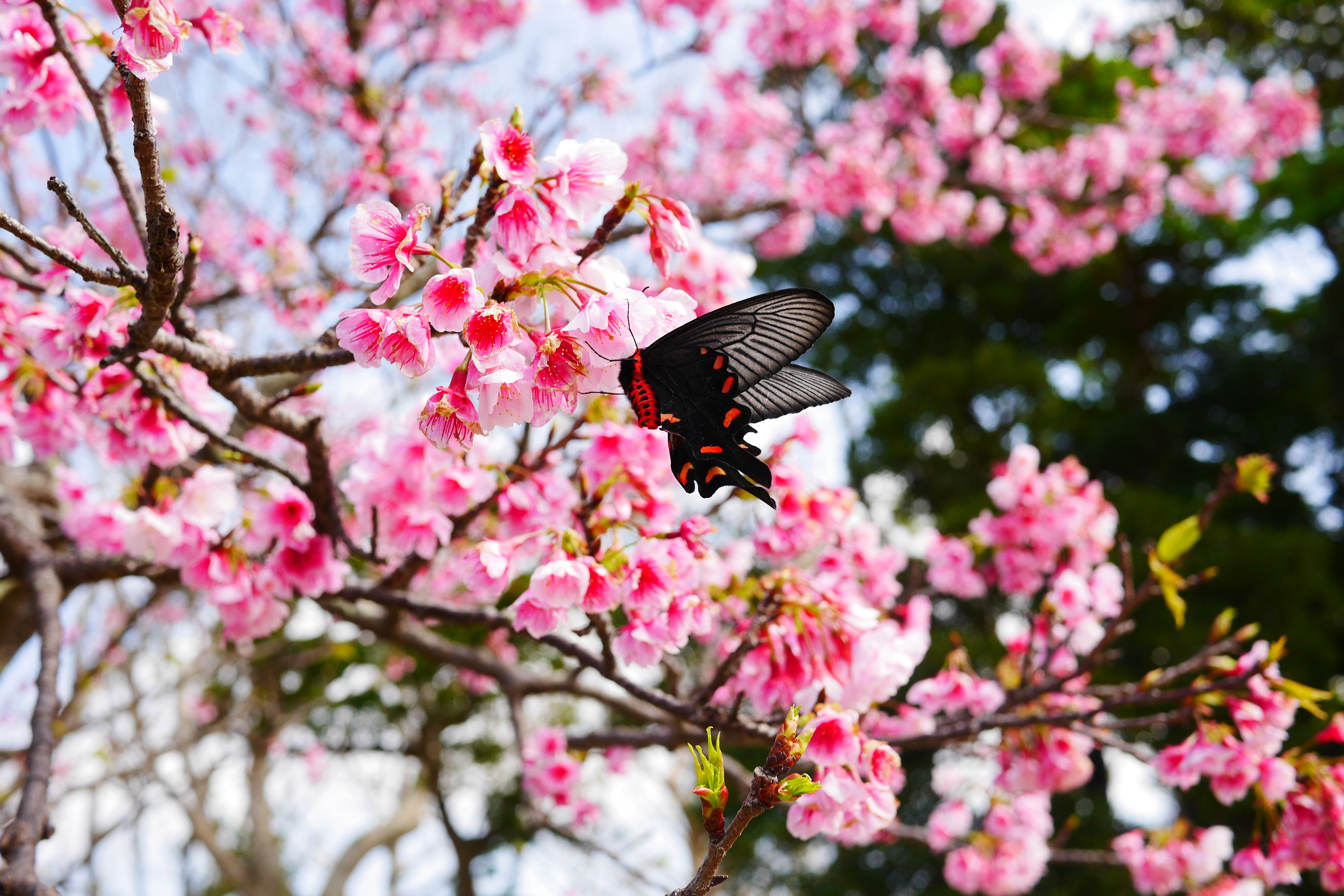
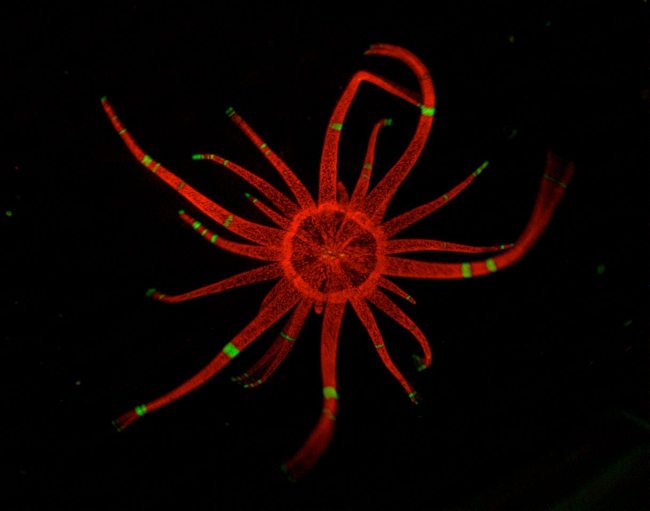
1. Oral disk (~1.5mm diameter) and tentacles of an Aiptasia pallida anemone. A. pallida tissue contains symbiotic algae called zooxanthellae which photosynthesize and provide nutrients for their host anemone. Zooxanthellae autofluoresce when excited, and appear red in this image. Green banding on the tentacles is tissue expressing GFP, fluorescing green here. As the image was taken, the anemone began to wake up from anesthesia and started waving its tentacles around, the motion of which can be seen at the tips of the tentacles. Image taken with with CCD camera attachment to Leica MZ16F fluorescence dissection scope using GFP filter.

Image

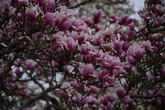
2. This is a photograph of a magnolia tree in the Ben West parking lot, taken on a rainy spring day in 2012.

Image

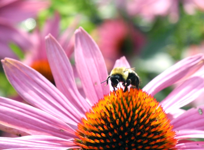
3 This photo was taken with a Nikon P7000 two summers ago during a family vacation at the Thuya Gardens near Northeast Harbor, Maine. The subject is a honey bee pollinating a flower. When the photo was taken, late August, the garden was in full bloom and the air was literally full of honey bees. It took several attempts and a great deal of patience, but I was finally able to get a non-blurry closeup of a bee.


Image

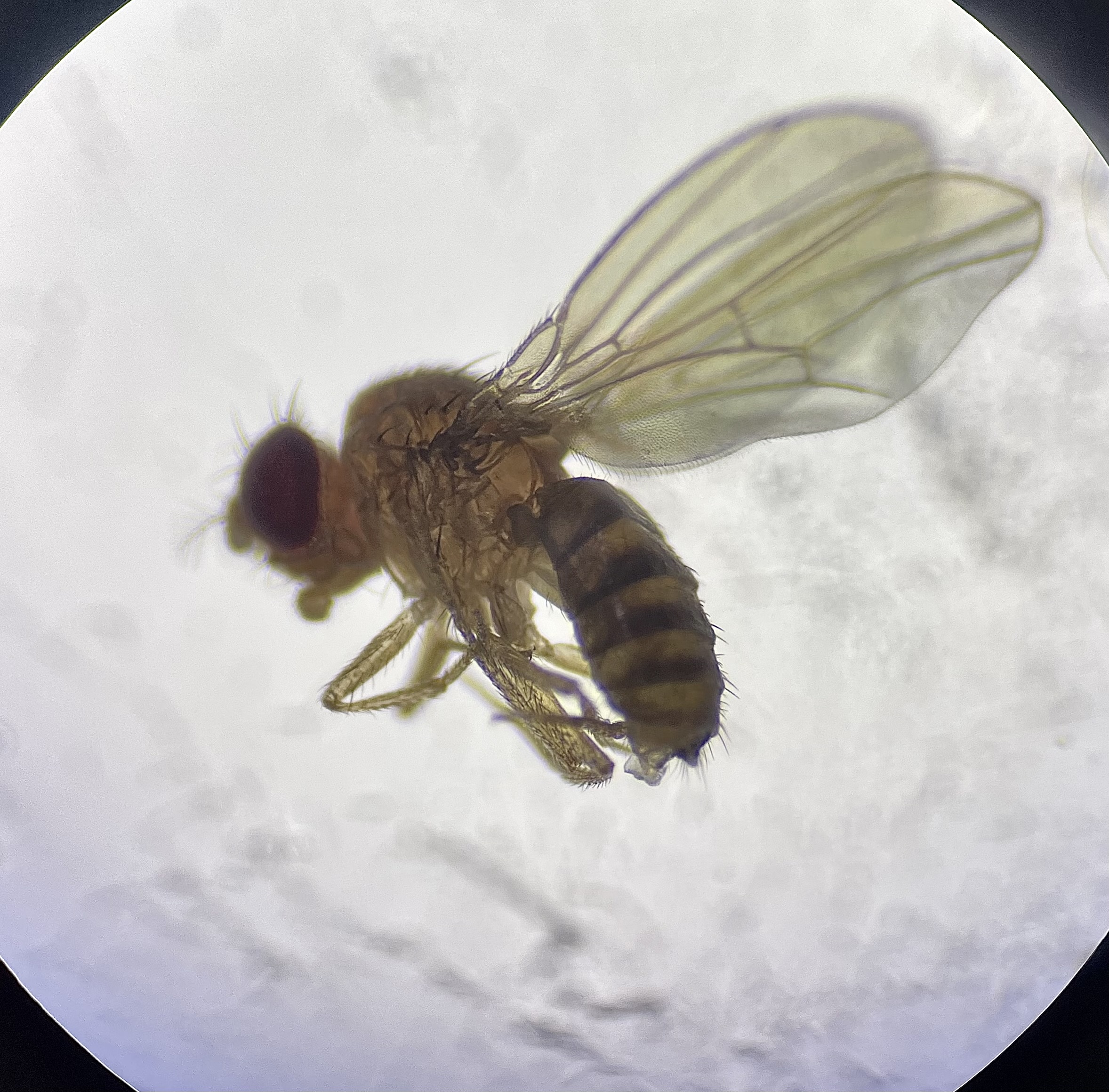
4 I took this photo during a brightfield microscopy session in my cell bio lab. It features an anemone (aiptasia pallida) and its dinoflagellate symbionts.


Image

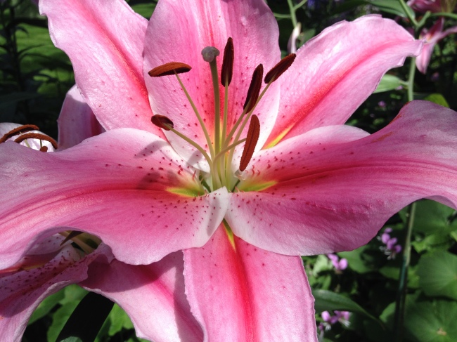
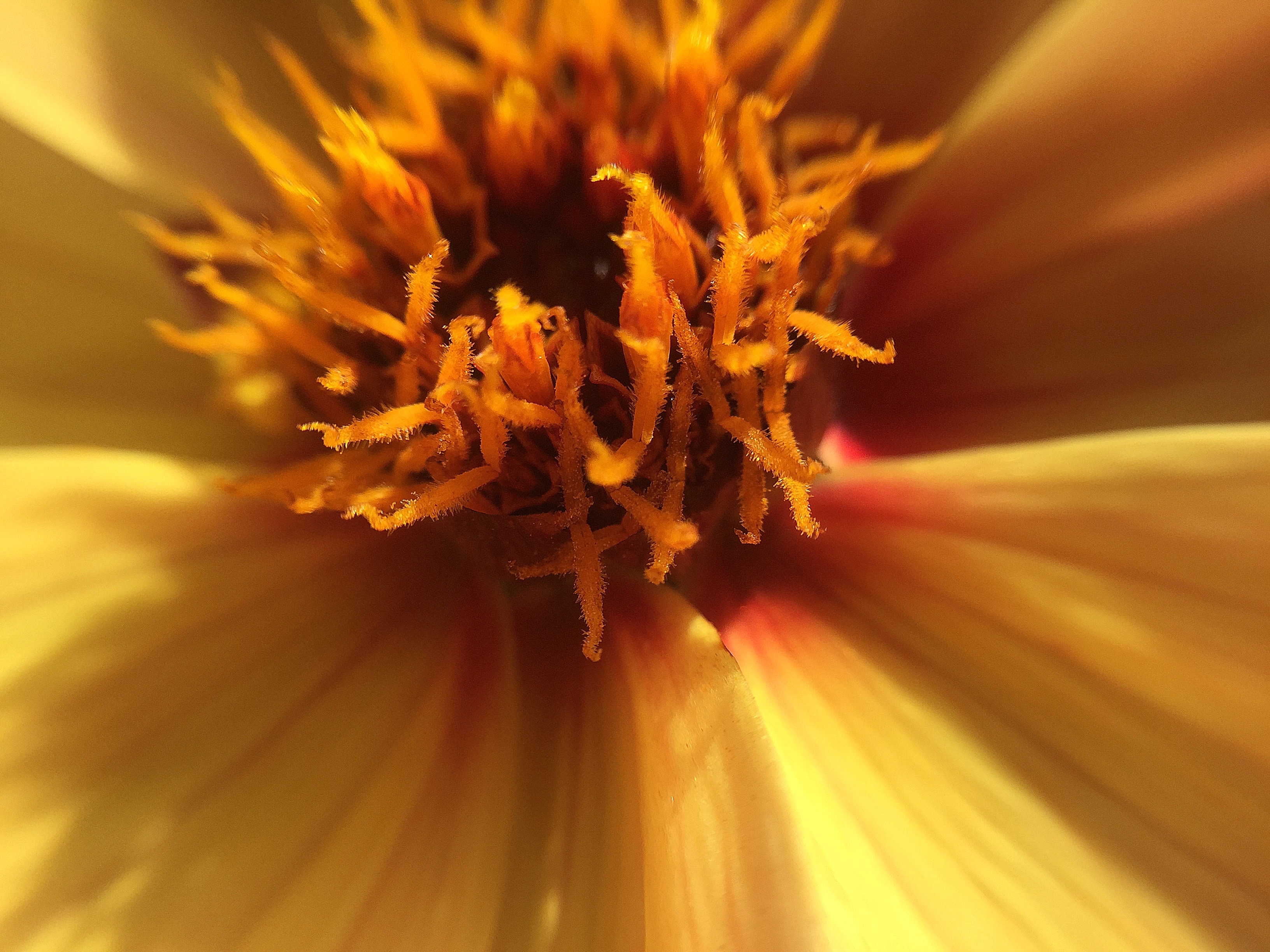
5 This is a Stargazer Lily, a type of oriental hybrid of lily, found at Longwood Gardens. This plant is an angiosperm (flowering plant) and this image shows the stamen, containing pollen, and the stigma, which receives the pollen and leads to the ovaries. These lilies produce a beautiful smell and contain elaborate coloring, likely to attract pollinators to the flower to pollinate the plant.


Image

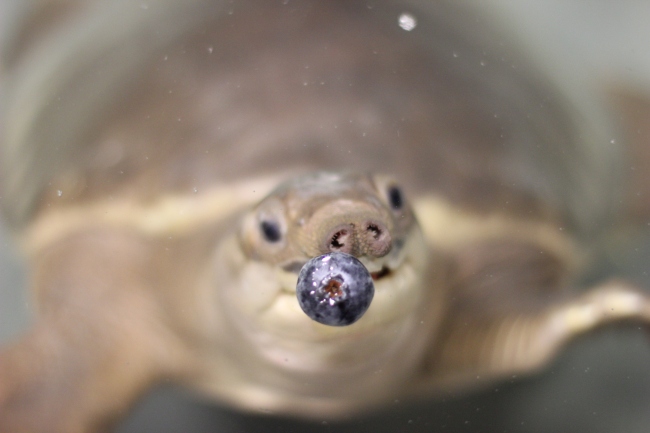
6 An Australian Pig-nosed turtle, Carettochelys insculpta, examines a blue berry. Pig-noses are the last extant members of the genus Carettochelys. Found only in Australia and New Guinea, Pig-noses are very different from other species of turtles. Their name comes from their pig-like nostrils, which act like snorkels for the aquatic turtle and house sensory organs to aid with hunting. The Pig-nosed turtle has full flippers resembling those of sea turtles rather than the webbed feet of its freshwater counterparts and is an agile swimmer. Its carapace is soft and leathery with a skin-covered plastron below.


Image

7 This image captures the textural depth and iridescence of the nacre-coated inner shell of Haliotis (Abalone), a marine gastropod mollusk. Colloquially known as “mother of pearl,” nacre is secreted by the Abalone’s epithelial cells and acts as a mechanism of defense, entombing invaders such as parasites and debris in a process called encystation. The iridescence characteristic of nacre stems from light diffraction and interference.

Image

8 A sea anemone specimen, order Actiniaria, at the Birch Aquarium, San Diego, California. Actiniaria lack the medusa phase of many other Cnidarians, but do posses the nematocysts (stinging cells) characteristic of the phylum.

9SY

9 The surface of the leaf of a Delosperma floribunda ‘Stardust’ plant, a succulent native to southern Africa. The translucent beads on the surface of the leaf are not drops of water – rather they are small liquid-filled globules that are part of the plant and likely act as lenses to focus light onto the chloroplasts that are visible directly underneath. This image was taken with a brightfield microscope.


Image

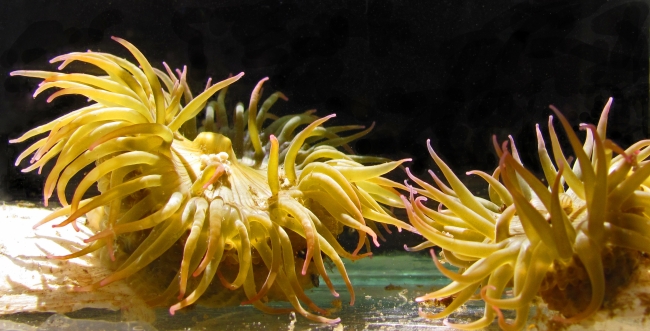
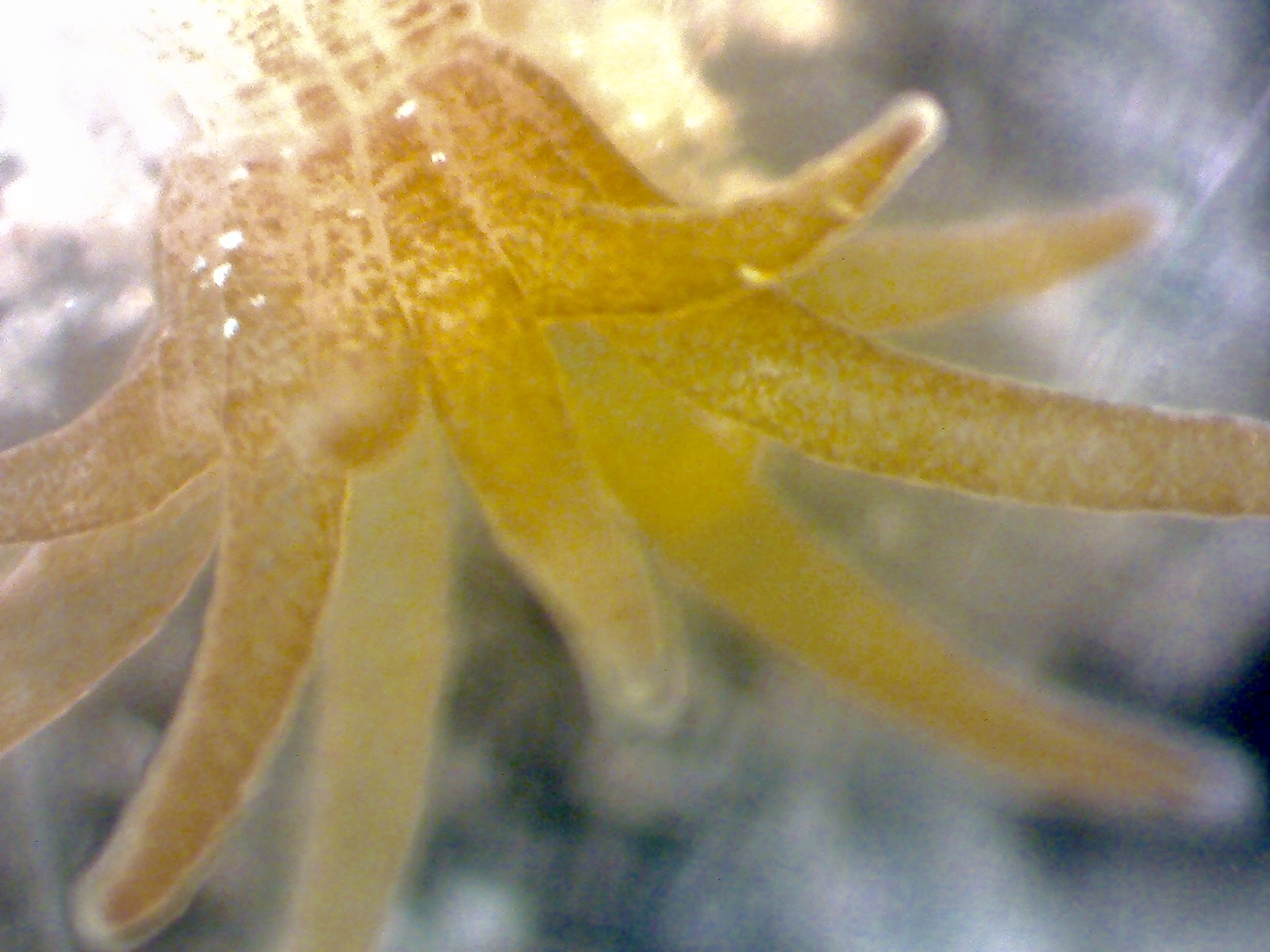
10 This is an image I captured in the summer of 2013 at Friday Harbor Laboratories of two polyps of the sea anemone Anthopleura elegantissima. My research is focused on the aggressive response that this clonal anemone mounts against nonclonemates. In this image, I have placed two nonclonemates together in a small aquarium and we can see their feeding tentacles beginning to touch. This tentacular stimulation can incite an anemone to either retreat or mount an aggressive response by inflating the specialized battle tenatcles, the acrorhagi, that are densely-packed with toxic nematocysts.

11AC

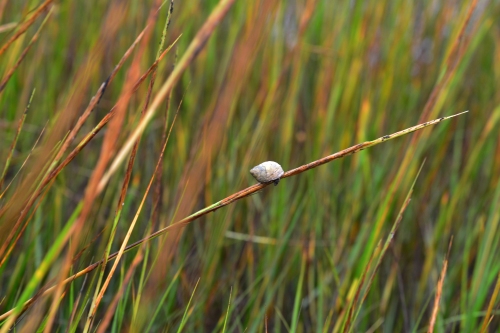
11 Littorina irrorata (marsh periwinkle) on spartina (cord grass) taken on Shackleford Banks, North Carolina. Though quite tiny, this sea snail is fascinating since it is believed to be a fungus farmer. With its radulla, this snail scratches the algae on the plant stem and then defecates in this area, encouraging the growth of fungi. This fungi is the main source of food for littorina.
ImageJ=1.48s unit=mm

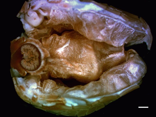
12 This image displays the inside of the opened seed of ipomoea alba, commonly known as the Moon Vine. This garden vine is most well known for its large white flowers which open only in the evening. The seed is marked by its thick seed coat surrounding tightly wrapped cotyledons, which will become the first leaves of the plant. This photo was taken with a dissecting scope. The scale bar marks a length of 1 millimeter.
13ST

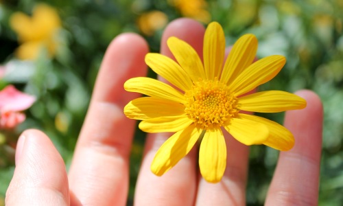
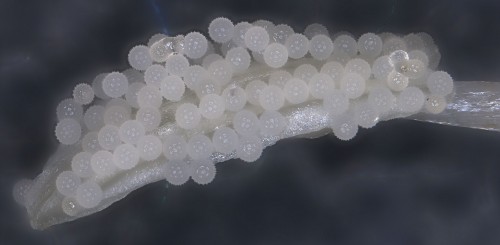
13 This photo depicts a Euryops pectinatus from Longwood Gardens in my palm.

14HRL_copy

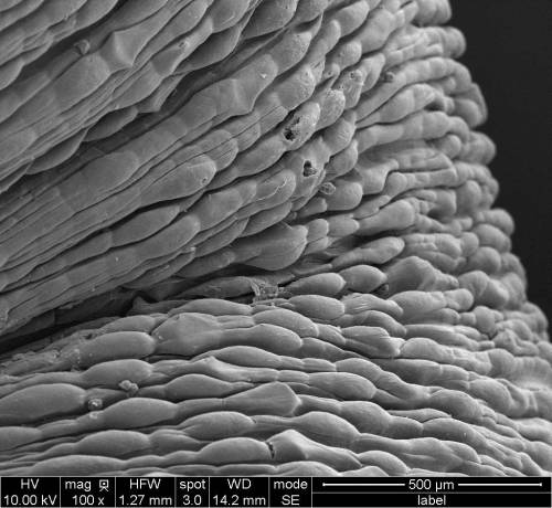
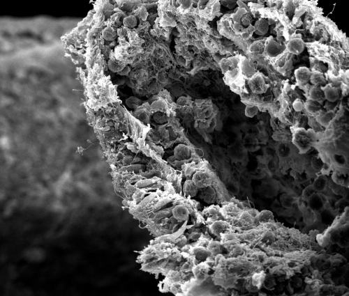
14 This is a picture of the surface of Leptosynapta clarki obtained with a scanning electron microscope. L. clakri is a small burrowing sea cucumber found in False Bay, San Juan Island, Wa. Unlike a typical sea cucumber, L. clarki does not have tube feet and has a smooth body wall, which gives L. clarki a vermiform look.
15DKK

15 This is a photograph of an Aiptasia pallida anemone, as seen through a compound light microscope. Photo was taken with an iPhone camera.
16DK2

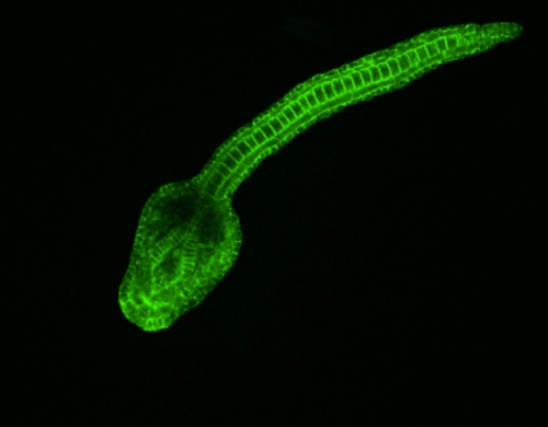
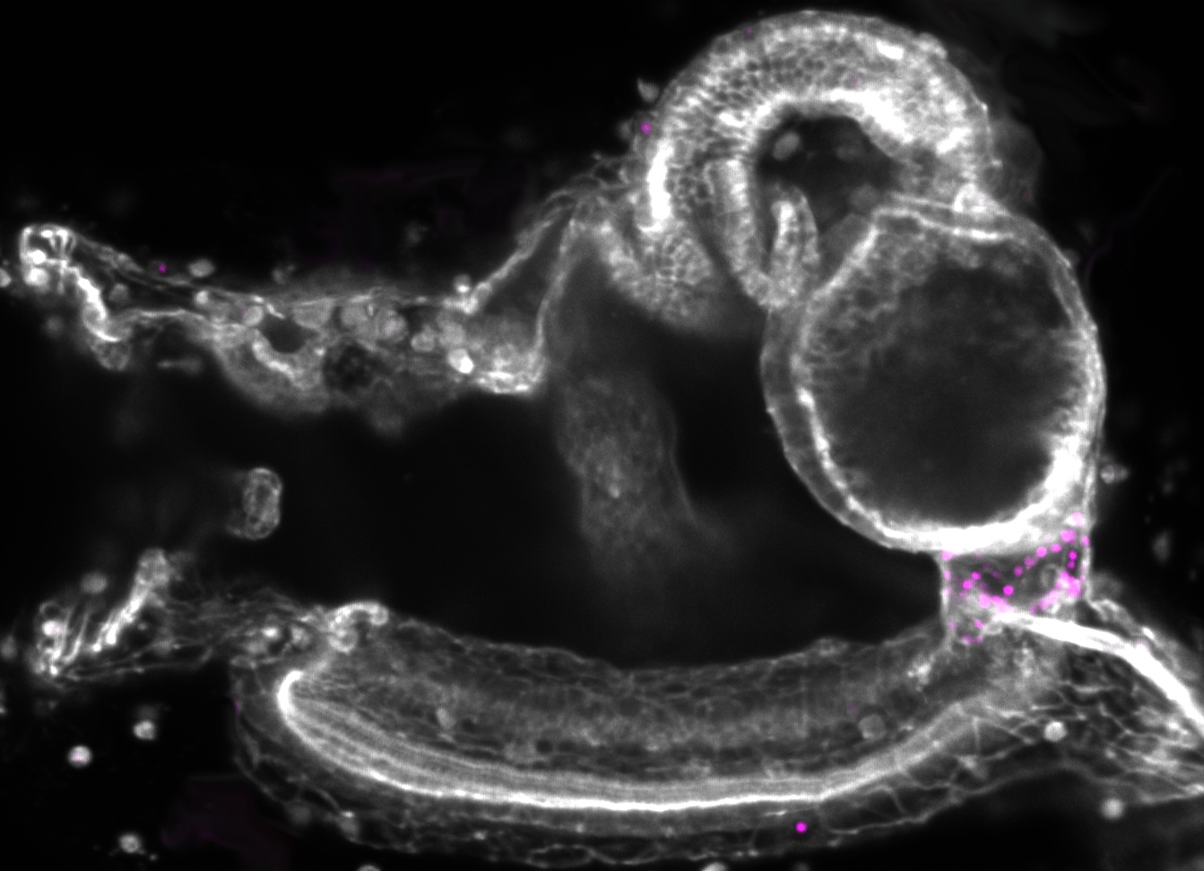
16 VIDEO: A Ciona intestinalis embryo 14 hours after fertilization, stained using a Phalloidin toxin. The embryo and its developing neural structures were viewed using confocal microscopy. (click here for video)

17AL

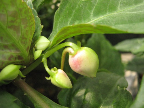
17 This Impatiens walleriana plant was grown as a morphology project for Plant Biology. Its new buds are just beginning to blush pink, foreshadowing the vibrant pink pigment that will accumulate in mature flowers. On the largest bud, the hooked nectary extends downward. It is from this nectary that the flower will secrete excess sugars in the form of sweet sugar water, which may attract pollinators (and pests!). Also visible in the foreground is the violet speckled underside of the Impatiens leaves. This contrasts with the relatively smooth green–interrupted only by a dusting of trichomes–of the top surface of the leaves.

18IB

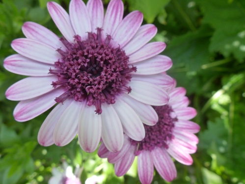
18 I took this picture when visiting Longwood Gardens on the 16th of April 2014 with my Biology 02 class. You can see beautiful little florettes in the middle of this purple and white flower.
19MS

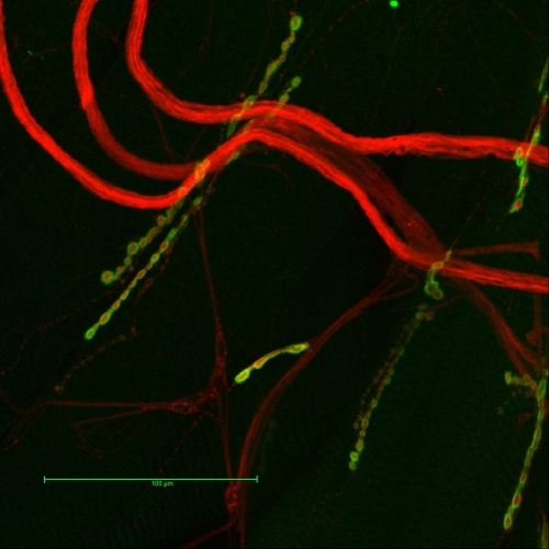
19 Confocal max projection of dorsal-side abdominal musculature treated with fluorescent Cy3 labeled anti-HRP (red), anti-DLG, and fluorescent Alexa488 labeled GAM (green). There appears to be six or seven NMJs labeled in the image, which have similar branching patterns of the presynaptic axon terminals innervating the muscles (appear parallel to one another). The postsynaptic membrane of the muscle envelops the motor neuron terminals, which is shown by the stereotyped “beads-on-a-chain” pattern of boutons. The boutons contain presynaptic neural tissue on the inside (red) with postsynaptic scaffold protein DLG on the outside (green). The boutons, which release glutamate, the primary excitatory transmitter at the larval NMJ, appear to be approximately the same size in each of the NMJs. Image is 20x objective with zoom factor 3. Scale bar = 100 mm.


20VG_copy

20 Cross section of a tentacle from Aiptasia pallida captured with electron microscopy. This image clearly shows the two dermal layers, the ectoderm and the endoderm, being separated by the mesoglea, a layer of extracellular matrix, along with the anemone’s endosymbiont, Symbiodinium.
21NC

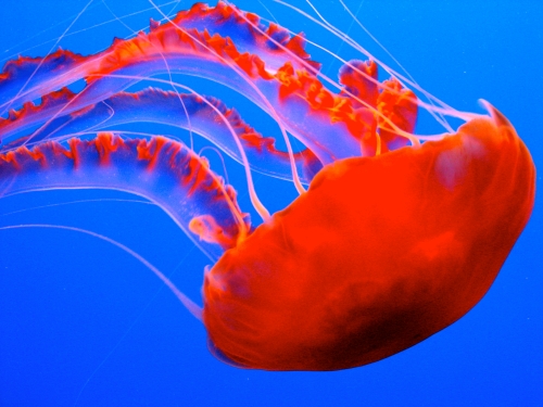
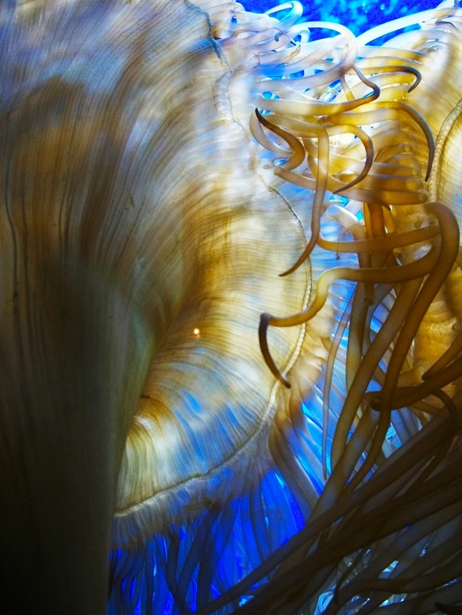
21 Sea Nettles (Chrysaora fuscescens) are native to the East Pacific Ocean, from Canada to Mexico. These jellyfish can grow to be 1 meter across, but they tend to remain smaller than 50cm. This photo was taken at the Monterey Bay Aquarium in California.
22CC

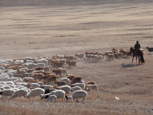
22 Symbiotic Relationships on the Steppe. My host father, a nomadic herder from Bayankhongor Aimag, uses an uurga to herd his sheep and goats in April. Mongolia’s continental climate poses a unique challenge to both nomads (who constitute roughly 30% of the country’s population) and herd animals with extreme high and low temperatures, as well as frequent droughts and dzuds (intense winter conditions). According to my host father, a changing climate is responsible for decreased precipitation, desertification, and the disappearance of many streams in his soum (county).

23CKC

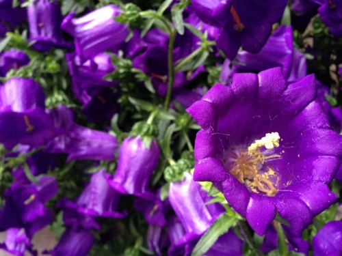
23 These are purple bell flowers taken by my iPhone 5.

24AB

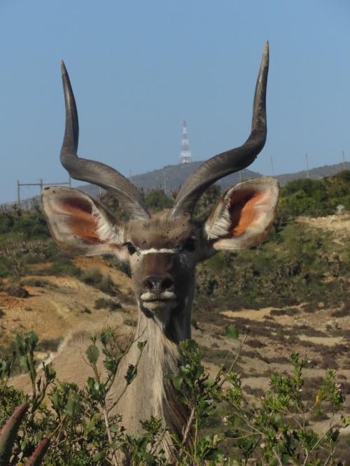
24 Male greater kudu (Tragelaphus strepsiceros)taken at the Garden Route Game Lodge in Albertinia, South Africa.  The greater kudu is a species of woodland antelope found throughout Eastern and Southern Africa.

25HA

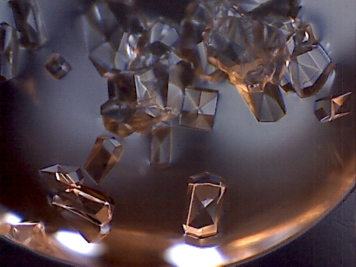
25 The image is a close up of CaCl2 salt crystals found in a single lysozyme drop. The crystals were synthesized using the hanging drop and vapor diffusion method, then photgraphed in the Biochemistry Lab of Swarthmore College using a digital QX5 microscope.

26BM

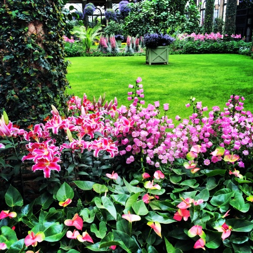
26 Green is good for the eyes, and a flower is a jubilant surprise.

27DM

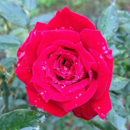
27 a rose from our rose garden here at Swarthmore. I took it the morning
after a rain and thought it looked good. I realize it isn’t the most scientific or biological
picture but I thought it wouldn’t hurt to submit it anyway.

Print

28 A 5µm section of the symbiotic anemone Aiptasia pallida. The specimen has been paraffin embedded, sectioned, and stained with Masson’s Trichrome Stain. The collagen is stained blue, the cytoplasm and muscle pink, and the nuclei black. The extracellular matrix, or mesoglea, is stained blue between the two cell layers, the ectoderm and gatroderm. In this particular section the hollow body column is filled with acontia, the threadlike defensive organs filled with explosive cnidocyte cells. The stained section was visualized using bright field microscopy.
29 CA

29 a photo I took of a sea anemone through the microscope!

30SW

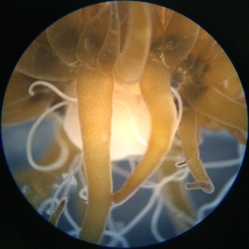
30 This is an anther from an Ipomoea purpurea (Morning Glory) flower of the ‘President Tyler’ variety in full bloom. This image was taken with an extended depth-of-field microscope on a glass slide.

31BN

31 VIDEO: Captured here are two species of bees collecting nectar. Some of these bees are true pollinators–they feed through the natural opening of the flower and potentially gather or unload pollen. Other bees pictured are “nectar robbers”, and instead extract nectar from a hole they poke in the base of the flower. This can allow them to feed on a wider variety of flowers, but generally does not result in pollination. (click here for video)

32AK

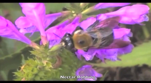
32 Three Italian honey bees drinking honey water. The picture was taken inside one of the Environmental Studies research project bee hives.

33JZ

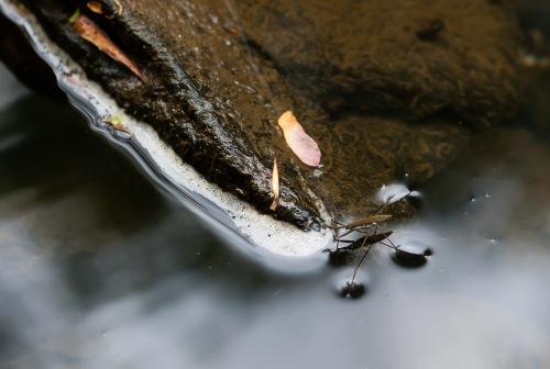
33 This is a member of the Gerridae family of bugs, more commonly known as water striders or water bugs. These bugs have more than one thousand hydrophobic microhairs per mm on their body, protecting them from splashes of water and giving them their namesake ability to walk on water. This one was found during springtime in the Crum.

34AK

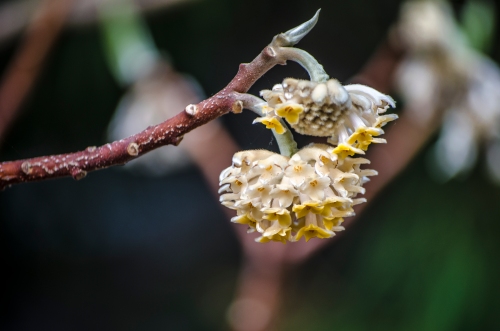
34 Oriental paperbush (Edgeworthia chrysantha) photographed on Swarthmore College’s campus. The plant’s bark is a source of high quality paper which gives the plant its English name, paper bush.

35JC

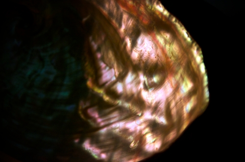
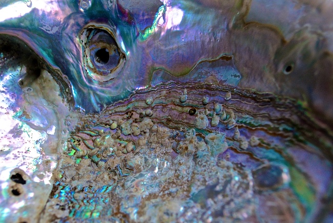
35 Shown here is a detail of the interior of an Abalone shell. The iridescent pattern is produced by the nacre, an organo-mineral composite comprised primarily of alternating layers of aragonite pseudohexagonal tablets and organic matrix secreted by the epithelial cells of the mantle tissue. Nacre coats the interior shell of bivalves, gastropods, and cephalopods and serves as a defense against parasites and debris.

36KB


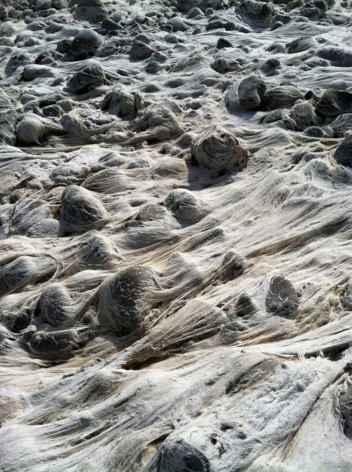
36 California is depleting its water sources at an unsustainable rate.” The most piercing, motivating thing can become dull when you hear it over and over. We still have to prompt ourselves, in a different vocabulary. This image was taken during a river clean-up effort of the Ventura River over spring break 2014, in a place where the rapid current once incessantly pulled at the now ghostly algae threaded around the rocks.


37EC_copy

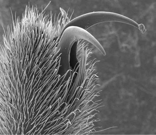
37 The front leg of a water strider from Crum Creek. Water striders are slender, dark colored insects of the family Gerridae, that can be seen on the surfaces of ponds, streams, and marshes throughout the Northern Hemisphere. Water striders’ hairy legs increase the total amount of displaced fluid by increasing the surface area that makes contact with the water. The claws of the front legs, shown in this picture, allow the water strider to dig into the water and pull themselves across the surface. This picture was taken with a scanning electron microscope at 120x magnification.

38GR

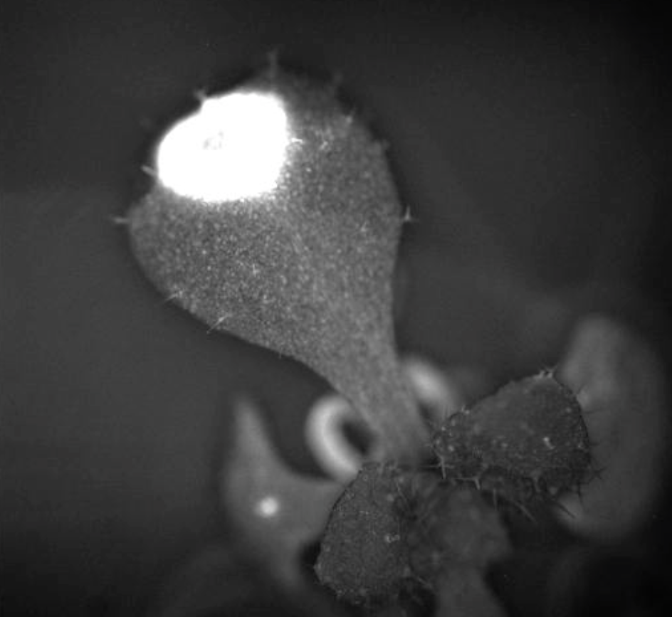
38 Hsp17.6 expression in 37°C heat shocked Arabidopsis root. The RootScope, an automated fluorescent microscope built by the Kaplinsky lab, was used to collect images and quantitate Hsp17.6p:GFP response in Arabidopsis seedlings to measure heat shock response over time.

Collage of 2013 submissions


Pages

Blog Stats

  • 22,001 hits

Enter your email address to subscribe to this blog and receive notifications of new posts by email.

Join 4 other subscribers